Processing Samples for Plant-Parasitic Nematodes

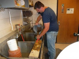
Figure 1: Processing a soil sample for nematodes
Figure 1: Processing a soil sample for nematodes.

Plant-parasitic nematodes are microscopic animals. The longest plant-parasitic nematode found in MI measures about 6 mm (ca. 1⁄4 inch) in length and that is the corn needle nematode. Female cyst nematodes achieve large enough sizes where they can be seen with the naked eye attached to roots on which they are feeding. However, to identify them to species they would need to be examined under a microscope or subjected to a molecular procedure such as PCR.

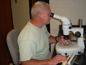
Figure 2: Fred Warner a Nematologist
Figure 2: Fred Warner – Nematologist

Virtually all plants have at least one nematode parasite. Economic losses on some crops plants can be significant due to feeding by nematodes. For this reason, it is important that nematode populations are monitored. In order to avoid or diagnose nematode problems, it is imperative soil and plant tissue samples are collected and then delivered/sent to a lab such as MSU Plant and Pest Diagnostics to assess nematode population densities. 

All plant-parasitic nematodes spend at least part of their life histories in the soil. To extract/ remove nematodes from the soil, we use a process called centrifugation/flotation. Nematodes, as well all other soil invertebrates, fungi, etc., are suspended in water and decanted over nested sieves (fig. 1). The sample is then spun in a centrifuge and all objects with higher specific gravities than water pellet in the bottom of the centrifuge tube. The water is then poured off after spinning and is replaced by a sucrose solution.

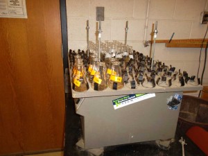
Figure 3: Flasks on Shaker

The pellet is re-suspended and the sample spun a second time. With this form of gradient centrifugation, nematodes, most other microscopic animals, fungi, weed seeds, etc., are suspended in the sucrose with only the sand grains forming a pellet. The sucrose solution is then poured over a fine-mesh sieve to catch the nematodes. They are then rinsed into a test tube and are ready to be identified and counted under a dissecting or inverted microscope (fig. 2).

Some nematodes enter plant tissues to feed. Most feed within or on root tissue but there are some species that feed on foliage. To extract nematodes from plant tissues, we use a shaker technique where roots or leaves are washed, weighed and then placed in flasks containing an incubation solution (fig. 3). After approximately 72 hours on the shaker, most of the nematodes have moved out of the plant tissues and into the solution. The solution is then poured over sieves trapping the nematodes. The nematodes are rinsed into test tubes and are once again ready for microscopic observation.

There are other procedures that can be used to assess nematode population densities but centrifugation/flotation and the shaker are by far the ones most commonly utilized at MSU Plant and Pest Diagnostics. The biggest advantage they offer is the speed in which nematode samples can be processed.

07.17.14

Comments are closed.

Other Documents in this Series

Accessibility Questions:

For questions about accessibility and/or if you need additional accommodations for a specific document, please send an email to ANR Communications & Marketing at anrcommunications@anr.msu.edu.